
Thymosin Alpha 1 Dosage: Chart, Timing and Mixing

What Is the Standard Thymosin Alpha 1 Dosage?
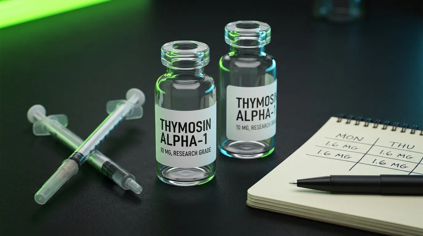
A pharmacy review gives the hepatitis B and C thymosin alpha 1 dosage as 1.6 mg under the skin twice a week. That equals 900 micrograms per square metre of body surface (Ancell et al. 2001). One hepatitis B trial treated patients for 6 or 12 months (Ancell et al. 2001). No cited source tested cycling.
This content is provided for educational purposes only. Thymosin alpha-1 is a research compound in most jurisdictions. A qualified clinician can advise on any health decision.
Thymosin alpha-1 is also sold as thymalfasin or Zadaxin. TA-1 is the short form used below.
Here is that review's hepatitis schedule in one place.
- Dose per injection: 1.6 mg subcutaneous, which means under the skin.
- Frequency: twice weekly.
- Duration: One hepatitis B trial treated patients for 6 or 12 months. No cited source tested cycling.
- Injection site: under the skin. The cited sources name no body site.
Thymosin Alpha 1 Dosage Chart: What Did the Trials Actually Use?
Both trials below used the same 1.6 mg injection. What changed was how often, and for how long.
| Trial / context | Dose | Frequency | Duration | Primary outcome |
|---|---|---|---|---|
| Hepatitis C, with interferon (Sherman 1998) | 1.6 mg SC | Twice weekly | 26 weeks | 37.1% ALT normalisation vs 16.2% interferon alone |
| Severe sepsis (Wu et al. 2013) | 1.6 mg SC | Twice daily x 5 days, then once daily x 2 days | 7 days | 28-day death rate 26.0% vs 35.0% control (P = 0.062) |
The hepatitis C row is worth setting out in full. The trial was randomised, double-blind and placebo-controlled. It randomised 109 patients with chronic hepatitis C. The combination arm received 1.6 mg of TA-1 under the skin twice weekly, alongside interferon-alpha 2b. Biochemical responders were followed for 26 weeks. At the end of treatment, ALT had normalised in 37.1% of the combination group. The interferon-alone arm reached 16.2% (Sherman et al. 1998). ALT is a liver enzyme that rises when liver cells are damaged.
For the vial maths, see the thymosin alpha 1 dosage calculator.
What Is the Thymosin Alpha 1 Dosage Per Day?
There isn't one, on the standard hepatitis schedule. The review above gives the dose twice a week, not daily. The sepsis trial below is the exception, with two injections a day at first.
The twice-weekly schedule surprises people who look at the pharmacokinetics first. Pharmacokinetics is how the body absorbs and clears a drug. TA-1 has a serum half-life of about two hours. Peak serum level comes within two hours, a time called Tmax. By 24 hours, circulating TA-1 is back to baseline (Ancell et al. 2001). Those are short kinetics by any measure.
So why would a twice-weekly schedule work at all? The sources cited here do not test that question directly. They do track immune markers over days, not hours. In a sepsis trial, HLA-DR on monocytes rose more with TA-1 than without, by day 3 and by day 7 (Wu et al. 2013). HLA-DR is a protein these white blood cells use to show germs to T cells.
| Parameter | Value |
|---|---|
| Tmax (time to peak serum level) | Within 2 hours |
| Serum half-life | ~2 hours |
| Return to baseline | Within 24 hours |
What Thymosin Alpha 1 Dosage When Sick or Acutely Immune Suppressed?
The best-tested acute setting is severe sepsis. A trial in six Chinese hospitals randomised 361 intensive care patients (Wu et al. 2013). The TA-1 group got 1.6 mg under the skin twice a day for five days. Then it was once a day for two more days.
Deaths from any cause within 28 days were 26.0% with TA-1 and 35.0% without. The authors call that P value of 0.062 marginal. A P value is a measure of how easily chance alone could explain a result. The usual cut-off for a firm result is 0.05. No serious drug-related side effect was recorded. TA-1 was given on top of standard sepsis care.
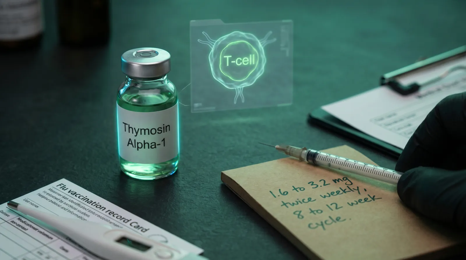
Vaccination is the other timing question. Older people respond less well to vaccines, which is why TA-1 was tested alongside them (Ershler et al. 2007). One double-blind trial enrolled 90 men aged 65 to 99. They got TA-1 or placebo with the 1986 flu vaccine (Gravenstein et al. 1989). The TA-1 dose was 900 micrograms per square metre under the skin, twice weekly for eight doses. No toxicity was seen in either group. A later review of these trials reports enhanced vaccine responses with TA-1 (Ershler et al. 2007). The timing detail sits in thymosin alpha-1 for post-viral illness, Lyme and vaccine response.
How Was a Vial Mixed for the Thymosin Alpha 1 Injection Dosage?
In the sepsis trial, the drug came as lyophilised, or freeze-dried, powder. It was mixed with 1 ml of the provided diluent, the liquid used to dissolve it. That diluent was sterile water for injection. The final concentration of TA-1 was 1.6 mg/ml (Wu et al. 2013).
A certificate of analysis, or COA, is a lab report on a batch's identity and purity. How to read a peptide COA walks through what one should say.
What Is the Thymosin Alpha 1 Cancer Dosage in Trials?
The largest cancer trial cited here randomised 488 people with metastatic melanoma (Maio et al. 2010). Metastatic means the cancer had spread. TA-1 was tested at 1.6 mg, 3.2 mg or 6.4 mg, added to the chemotherapy drug dacarbazine. Four of the five groups also got interferon-alpha. The abstract does not give the injection schedule.
The two 3.2 mg groups had 10 and 12 tumour responses, against four in the control group. Adding TA-1 caused no extra toxicity (Maio et al. 2010). So in this trial, the cancer dosage was not fixed at 1.6 mg.
How Does Thymosin Alpha 1 Work at the Receptor Level?
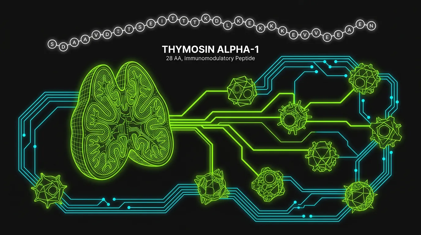
TA-1 is a 28-amino acid peptide. It comes from thymosin fraction 5. Allan Goldstein first described it in 1972 (Pierluigi et al. 2010).
Dendritic cells are immune cells that alert T cells to a threat. TA-1 activates one type, plasmacytoid dendritic cells, through TLR9 (Romani et al. 2007). Toll-like receptors are pattern-recognition proteins on innate immune cells. They evolved to spot the signatures of bacteria and viruses. TLR-9 sits inside the cell, in the endosome, a small sac holding material the cell has taken in. It detects a DNA pattern called unmethylated CpG, typical of microbial genomes.
In mouse dendritic cells, the signal runs through MyD88, an adaptor protein that passes it inward. It then runs through p38 MAPK and NFkB (Romani et al. 2004). p38 MAPK is a stress-signalling enzyme. NFkB is a switch protein that turns on immune genes. The dendritic cells matured and made IL-12, a messenger that steers T cells (Romani et al. 2004). A study in human dendritic cells found p38 MAPK and NFkB switched on quickly too (Yao et al. 2007).
In plain terms, TA-1 makes dendritic cells better at their job of instructing T cells. In human cell culture, TA-1 significantly upregulated CD80 and MHC class I and class II. These are surface proteins a dendritic cell uses to show antigen to a T cell. The dendritic cells were grown from blood monocytes. Thymosin beta-4 did not do this (Yao et al. 2007).
CD80 and CD86 are signals a T cell needs alongside the antigen. Without them, a T cell meeting antigen becomes anergic, which means switched off rather than switched on. In mice, TA-1 switched on a Th1 response against a fungus (Romani et al. 2004). Th1 cells are helper T cells that make IFN-gamma, IL-2 and TNF-alpha. These are cytokines, the immune system's chemical messengers.
The same peptide can also calm the immune system. The sepsis trial report notes that TA-1 can raise IL-10 and the share of regulatory T cells (Wu et al. 2013). IL-10 is a calming messenger, and regulatory T cells damp inflammation. In animal studies, TA-1 also switched on an enzyme called IDO in dendritic cells, which builds tolerance (Romani et al. 2007). Tolerance means the immune system stands down instead of attacking. One review asks whether this makes TA-1 a regulator of regulators. It is not a simple immune stimulant (Pierluigi et al. 2010).
The thymus link explains the name. TA-1 was first isolated as the compound that restored immune function in animals with no thymus (Tuthill et al. 2010). In the body, it is cut from a larger protein, prothymosin alpha, in many tissues (Romani et al. 2007). Thymic output falls sharply after puberty and keeps falling. That is part of immunosenescence, the age-related decline in immune function.
Hormones are a separate question. The sources cited here do not measure cortisol, testosterone, estrogen, GH or IGF-1 in people on TA-1, so this page makes no claim about them.
Does Thymosin Alpha 1 Dosage Change When You Stack It?
The sources cited here tested TA-1 with only two partners. The hepatitis C trial used the usual 1.6 mg twice weekly alongside interferon-alpha (Sherman et al. 1998). The melanoma trial added 1.6, 3.2 or 6.4 mg to chemotherapy (Maio et al. 2010). No cited source tested TA-1 with the other compounds below.
TA-1's studied actions are on immune cells. Whether it clashes with, or adds to, other compounds is untested in these sources.
Interferon-alpha (IFN-alpha) is an immune protein used as an antiviral drug. In the hepatitis C trial above, adding TA-1 to it raised the end-of-treatment ALT response from 16.2% to 37.1%.
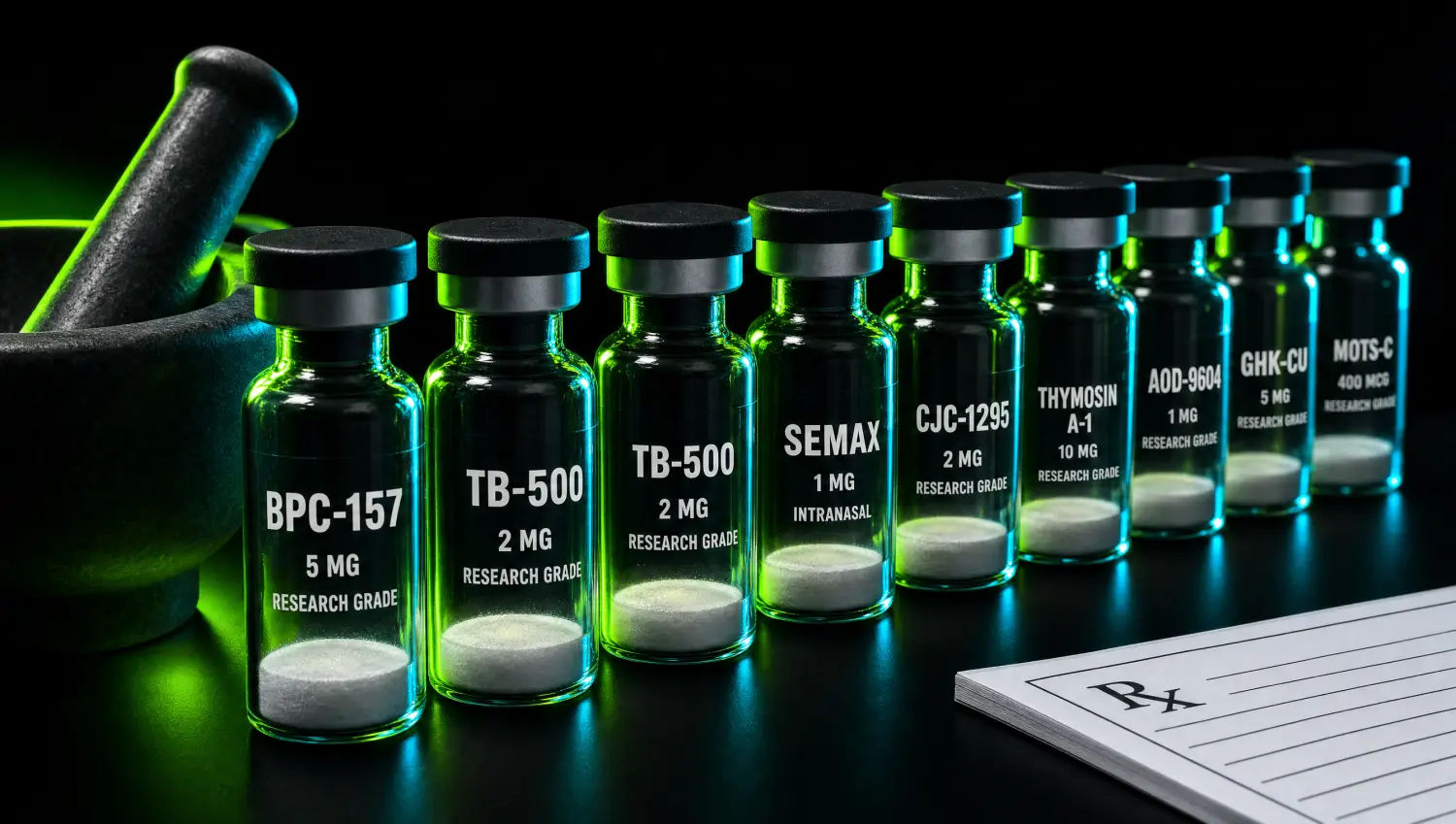
TB-500 deserves a separate note, because the naming confuses people. Thymosin beta-4 and thymosin alpha-1 both come from thymosin fraction 5. They are different peptides with different targets. TA-1 acts on immune cells. Thymosin beta-4 is a 43-amino acid peptide. It is the main protein that holds single units of actin, a building block of the cell's inner frame. It plays a key part in skin and eye wound healing (Goldstein et al. 2005). The TB-500 complete guide covers that side. For the broader logic of combining peptides, see peptide stacks that work, and for a gut-repair pairing, BPC-157 gut health.
What Should You Monitor on a Thymosin Alpha 1 Dosage Protocol?
Each cited trial tracked a small set of markers. The table lists what each one measured and found. None of them sets a monitoring plan for use outside a trial.
| Marker | Trial | What was reported |
|---|---|---|
| ALT, a liver enzyme | Hepatitis C, with interferon (Sherman 1998) | Normal at end of treatment in 37.1% on the combination vs 16.2% on interferon alone |
| HCV RNA, the hepatitis C viral load | Hepatitis C, with interferon (Sherman 1998) | Below baseline at weeks 8, 16 and 24 on the combination |
| HBV DNA, the hepatitis B viral load | Hepatitis B trial (Ancell 2001 review) | Cleared in 40.6% after 6 months and 25.6% after 12 months of TA-1, vs 9.4% of untreated controls |
| HLA-DR expression on monocytes | Severe sepsis (Wu et al. 2013) | Rose more with TA-1 than without, by day 3 and day 7 |
| Flu antibody titre | Men aged 65 to 99 (Gravenstein 1989) | Response defined as a four-fold rise 3 to 6 weeks after the vaccine |
HLA-DR on monocytes is a research marker, not part of a routine blood count. How to vet a peptide supplier covers the testing standards that separate reliable sources from the rest.
Is Thymosin Alpha 1 Safe, and Where Does the Research Use Line Sit?
A pharmacy review calls TA-1 well tolerated. Most studies saw only local irritation at the injection site (Ancell et al. 2001). The sepsis trial recorded no serious drug-related side effect (Wu et al. 2013). The older men's flu trial saw no toxicity (Gravenstein et al. 1989). In the melanoma trial, adding TA-1 caused no extra toxicity (Maio et al. 2010).
TA-1 is sold as Zadaxin. It is used worldwide for hepatitis B and C, certain cancers and vaccine enhancement (Wu et al. 2013). Large phase-3 trials have been run in the United States, Italy and China (Tuthill et al. 2010). Where it is not a licensed medicine, it comes through compounding pharmacies or research peptide suppliers. Checking it falls on the buyer. Our recommended sources page lists suppliers with independently verified per-batch testing.
Outside formal clinical trials, any use of thymosin alpha-1 is research use. A qualified clinician can advise on any health decision. This article is educational and is not medical advice.
Where to buy Thymosin Alpha-1
The hard part with Thymosin Alpha-1 isn't the protocol. It's finding a supplier that can prove what's in the vial. We assessed dozens against per-batch, third-party testing. A handful passed.
See the sources that passed →References
- Pierluigi B, D'Angelo C, Fallarino F, et al. Thymosin alpha1: the regulator of regulators? Ann N Y Acad Sci 2010. Read the record
- Tuthill C, Rios I, McBeath R. Thymosin alpha 1: past clinical experience and future promise. Ann N Y Acad Sci 2010. Read the record
- Sherman KE, Sjogren M, Creager RL, et al. Combination therapy with thymosin alpha1 and interferon for the treatment of chronic hepatitis C infection. Hepatology 1998. Read the record
- Yao Q, Doan LX, Zhang R, et al. Thymosin-alpha1 modulates dendritic cell differentiation and functional maturation from human peripheral blood CD14+ monocytes. Immunol Lett 2007. Read the record
- Ancell CD, Phipps J, Young L. Thymosin alpha-1. Am J Health Syst Pharm 2001. Read the record
- Wu J, Zhou L, Liu J, et al. The efficacy of thymosin alpha 1 for severe sepsis (ETASS): a multicenter, single-blind, randomized and controlled trial. Crit Care 2013. Read the record
- Romani L, Bistoni F, Gaziano R, et al. Thymosin alpha 1 activates dendritic cells for antifungal Th1 resistance through toll-like receptor signaling. Blood 2004. Read the record
- Romani L, Bistoni F, Montagnoli C, et al. Thymosin alpha1: an endogenous regulator of inflammation, immunity, and tolerance. Ann N Y Acad Sci 2007. Read the record
- Gravenstein S, Duthie EH, Miller BA, et al. Augmentation of influenza antibody response in elderly men by thymosin alpha one. J Am Geriatr Soc 1989. Read the record
- Ershler WB, Gravenstein S, Geloo ZS. Thymosin alpha 1 as an adjunct to influenza vaccination in the elderly: rationale and trial summaries. Ann N Y Acad Sci 2007. Read the record
- Goldstein AL, Hannappel E, Kleinman HK. Thymosin beta4: actin-sequestering protein moonlights to repair injured tissues. Trends Mol Med 2005. Read the record
- Maio M, Mackiewicz A, Testori A, et al. Large randomized study of thymosin alpha 1, interferon alfa, or both in combination with dacarbazine in patients with metastatic melanoma. J Clin Oncol 2010. Read the record
Where to buy Thymosin Alpha-1
The hard part with Thymosin Alpha-1 isn't the protocol. It's finding a supplier that can prove what's in the vial. We assessed dozens against per-batch, third-party testing. A handful passed.
See the sources that passed →Share this article
Frequently Asked Questions
What is the standard dose of thymosin alpha 1 and why was it set at 1.6 mg?
Does thymosin alpha-1 affect hormones like testosterone or cortisol?
Can thymosin alpha-1 improve vaccine response in immunocompromised or elderly patients, and what dosing timing is recommended?
How long does thymosin alpha-1 take to work and what immune markers shift first?
What is the difference between thymosin alpha-1 and thymosin beta-4 (TB-500)?
Does thymosin alpha-1 need to be cycled or can it be used continuously?
Read Next
Disclaimer: This content is for educational purposes only. These compounds are intended for research use. Nothing here is medical advice. Always work with a qualified clinician before making changes to your health protocol.