
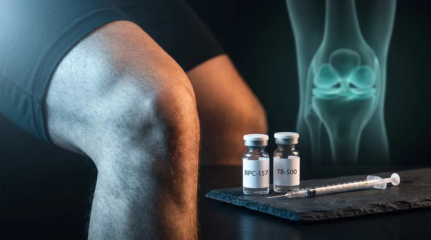
Best Peptide Stack for Torn Meniscus Without Surgery: BPC-157, TB-500, and Recovery Protocol (2026)
How BPC-157 and TB-500 Signal Meniscal Repair at the Molecular Level
BPC-157 drives meniscal repair by upregulating VEGF and GH receptors, accelerating collagen I/III cross-linking through fibroblast activation. TB-500 mobilises stem cells via actin-regulated cell migration and activates HIF-1α-dependent angiogenesis. Together, these two receptor-level pathways address the core vascular deficit that makes white-zone tears so resistant to healing.
The Core Biological Problem: Why the Meniscus Does Not Heal Itself
Before examining receptor pathways, it is worth understanding exactly why meniscal tissue occupies such a difficult position in connective tissue biology. The meniscus is a fibrocartilaginous wedge whose extracellular matrix is dominated by type I collagen arranged in circumferential bundles, with smaller amounts of type II collagen at the core. This architecture transfers compressive loads across the tibial plateau, distributing up to 70% of the knee's total contact force during weight-bearing. Makris et al. 2011 described the zonal vascular anatomy that defines its healing capacity: the peripheral red-red zone carries synovial vascularity and retains some regenerative potential, while the inner white-white zone is avascular, aneural, and almost entirely dependent on diffusion from synovial fluid for nutrient exchange.
That diffusion-based nutrition is sufficient for homeostatic maintenance but wholly inadequate for repair. Repair requires the delivery of progenitor cells, oxygen, and growth factors at concentrations orders of magnitude above basal levels. Without blood vessels penetrating the injury site, none of that occurs. Conservative physiotherapy, corticosteroid injections, and hyaluronic acid supplementation do not resolve this biology. They modulate symptoms. The vascular deficit remains.
This is not a peripheral detail. It is the central mechanistic reason why peptide researchers focus on angiogenesis as the primary target for meniscal regeneration. Both BPC-157 and TB-500 converge on neovascularisation through distinct but complementary receptor-level mechanisms, which is why their combination has attracted serious preclinical interest.
BPC-157: VEGF Upregulation, GH Receptor Sensitisation, and Collagen Remodelling
BPC-157, formally Body Protection Compound 157, is a synthetic 15-amino acid peptadecapeptide derived from a gastroprotective protein in human gastric juice. Its molecular formula is C62H98N16O22. Despite its short sequence, it interacts with at least three distinct receptor-level systems relevant to connective tissue repair.
VEGF and Angiogenic Signalling
The most mechanistically significant action of BPC-157 in fibrocartilage repair is its upregulation of vascular endothelial growth factor (VEGF) and its cognate receptor VEGFR2 (KDR). VEGF is the primary mitogen for endothelial cells and the principal driver of angiogenic sprouting. In avascular tissue zones, VEGF concentration is the rate-limiting factor for new vessel formation. BPC-157 elevates VEGF expression at injury sites, effectively rescuing angiogenic signalling in tissue that would otherwise remain VEGF-depleted.
Sikiric et al. documented this mechanism across multiple preclinical models, noting that BPC-157 activates the FAK-paxillin pathway downstream of VEGFR2, promoting endothelial cell migration and tube formation. Sikiric et al. 2018 The FAK (focal adhesion kinase) and paxillin signalling axis is a well-characterised integrin-associated pathway that governs how endothelial cells interpret extracellular matrix signals and respond by extending lamellipodia into the avascular zone. When BPC-157 activates this pathway, it is not simply stimulating growth factor production in a non-specific sense. It is engaging a precise intracellular cascade that instructs endothelial precursor cells to migrate directionally toward tissue hypoxia.
This matters for the meniscus specifically because the white-zone tear creates a local hypoxic pocket. Angiogenic rescue via the VEGF-FAK-paxillin axis is the mechanism by which that pocket could theoretically become vascularised, converting a white-zone tear's biology toward that of a red-zone tear with genuine healing capacity.
Growth Hormone Receptor Sensitisation
A second distinct mechanism involves the growth hormone (GH) receptor axis. BPC-157 has been shown to upregulate GH receptor expression in musculoskeletal tissue, effectively amplifying the anabolic signal from endogenous GH without altering circulating GH levels. This is a receptor sensitisation effect, not a secretagogue effect. The distinction is clinically important: BPC-157 does not raise serum IGF-1 in the manner that GH secretagogues do, but it does appear to potentiate tissue-level GH signalling, particularly in fibroblasts and chondrocytes.
Fibroblast responsiveness to GH is directly relevant to collagen synthesis. GH receptor activation in fibroblasts stimulates the JAK2-STAT5 pathway, which drives transcription of collagen type I and type III genes. Staresinic et al. 2010 demonstrated in a rat Achilles and medial collateral ligament transection model that BPC-157 treatment produced statistically significant increases in type I collagen content at the repair site compared to controls, along with improved biomechanical load-to-failure values. The histological sections showed more longitudinally organised collagen fibres rather than the disorganised whorling characteristic of scar tissue.
Collagen organisation is not an aesthetic consideration. The mechanical properties of fibrocartilage, including stiffness, tensile strength, and fatigue resistance, are directly determined by the regularity of collagen fibril cross-linking. Poorly organised collagen, the kind typically deposited in avascular healing, is mechanically inferior. BPC-157's apparent ability to promote structured collagen deposition via GH receptor sensitisation addresses this problem at the biosynthetic level.
Anti-Inflammatory Cytokine Modulation
BPC-157 also modulates the NF-kB pathway, a master transcriptional regulator of pro-inflammatory cytokine production. In inflammatory environments, NF-kB drives the expression of IL-6, TNF-alpha, and IL-1beta, all of which are catabolic to connective tissue and inhibitory to fibroblast-mediated repair. BPC-157 downregulates NF-kB activity, reducing the local cytokine burden at injury sites. A 2024 systematic review encompassing 544 screened articles and 36 studies meeting inclusion criteria confirmed that BPC-157 consistently reduces inflammatory cytokines and improves structural and biomechanical outcomes across muscle, tendon, ligament, and bone injury models. Sikiric et al. 2024
The NF-kB suppression effect creates what might be described as a permissive microenvironment for repair: inflammation sufficient to recruit repair cells, but reduced enough that the catabolic cytokine tide does not overwhelm early fibroblast and chondroblast activity. This represents a qualitatively different mechanism from corticosteroid anti-inflammatory action, which is broadly immunosuppressive and inhibits both destructive and reparative processes.
For a practical introduction to dosing considerations, see our detailed guide on BPC-157 dosing protocol for musculoskeletal injuries.
TB-500: HIF-1α, Actin Dynamics, and Stem Cell Mobilisation
TB-500 is a synthetic analogue of thymosin beta-4 (Tb4), a naturally occurring 43-amino acid peptide encoded by the TMSB4X gene. It is ubiquitously expressed across virtually all nucleated cell types and is among the most abundant intracellular peptides in mammalian biology, present at concentrations of 200-500 micromolar in platelets and 10-100 micromolar in most other cell types. Its primary biological role is the sequestration of G-actin monomers, regulating the equilibrium between globular and filamentous actin and thereby controlling cytoskeletal dynamics.
Actin Regulation and Cell Migration
The connection between actin regulation and tissue repair is more direct than it might initially appear. Cell migration, the process by which repair-competent cells travel from their origin to the injury site, is entirely dependent on dynamic actin remodelling. A migrating cell extends lamellipodia (flat actin-rich protrusions) at its leading edge and retracts at its trailing edge. This requires precise, coordinated polymerisation and depolymerisation of actin filaments, exactly the process that thymosin beta-4 and its analogue TB-500 regulate.
By modulating the G-actin:F-actin ratio, TB-500 creates conditions that favour sustained cell migration. Sosne et al. demonstrated in corneal epithelial injury models that thymosin beta-4 accelerates wound closure by promoting actin organisation at the leading edge of migrating epithelial sheets. Sosne et al. 2002 While the tissue context differs from fibrocartilage, the actin migration mechanism is conserved across cell types. Fibroblasts, chondrocytes, and endothelial progenitors migrating into a meniscal tear zone all depend on the same cytoskeletal machinery.
HIF-1α Activation and Hypoxic Response
Hypoxia-inducible factor 1-alpha (HIF-1α) is a transcription factor that coordinates the cellular response to low oxygen conditions. In avascular tissue zones, HIF-1α is stabilised (it is normally rapidly degraded under normoxic conditions by prolyl hydroxylase enzymes) and drives the transcription of dozens of hypoxia-response genes, including VEGF, erythropoietin, glucose transporters, and several matrix metalloproteinases involved in remodelling.
TB-500 has been shown to activate HIF-1α signalling pathways, essentially amplifying the tissue's own hypoxic distress signal. Smart et al. demonstrated that thymosin beta-4 promotes cardiac progenitor cell survival under hypoxic conditions through HIF-1α-dependent mechanisms, with downstream activation of VEGF and anti-apoptotic gene expression. Smart et al. 2007 In the context of an avascular meniscal tear, HIF-1α activation via TB-500 would theoretically amplify the angiogenic signal already initiated by BPC-157's VEGF upregulation, creating a synergistic neovascularisation stimulus.
This HIF-1α-VEGF axis represents the molecular basis for the often-cited claim that TB-500 and BPC-157 are synergistic. They are not redundant; they activate the same angiogenic endpoint through different upstream inputs. BPC-157 drives VEGF from the growth factor receptor side; TB-500 amplifies the hypoxic transcriptional program that independently elevates VEGF and other repair-permissive genes.
Stem Cell Mobilisation via SDF-1/CXCR4
Perhaps the most remarkable mechanism attributed to thymosin beta-4 and its analogues is the mobilisation of CD34+ progenitor cells and mesenchymal stem cells (MSCs) from bone marrow to peripheral injury sites. This occurs through the SDF-1 (stromal cell-derived factor 1) and its receptor CXCR4 chemokine axis. Injury-damaged tissue releases SDF-1, which creates a chemotactic gradient that CXCR4-expressing progenitors follow.
TB-500 upregulates CXCR4 expression on progenitor cells, increasing their sensitivity to the SDF-1 gradient emanating from injury sites. Crockford et al. in their review of thymosin beta-4's cardiac repair mechanisms described this stem cell mobilisation as a key component of the peptide's regenerative activity in ischaemic tissue. Crockford et al. 2010 For meniscal repair, this mechanism is significant because MSCs can differentiate into chondrocytes, the cells responsible for fibrocartilage matrix production. Increasing MSC recruitment to the tear site does not simply accelerate existing repair processes; it potentially changes the cellular composition of the repair tissue itself.
For a comprehensive overview of TB-500's mechanisms across injury types, our TB-500 complete guide provides additional mechanistic context.
Integrin Pathways: How These Peptides Interface with the Extracellular Matrix
Both BPC-157 and TB-500 interact with integrin signalling, though through different mechanisms. Integrins are transmembrane receptors that span the cell membrane and connect the extracellular matrix (ECM) on the outside to the actin cytoskeleton on the inside. They function as bidirectional signal transducers: matrix stiffness, composition, and topography are sensed by integrins and converted into intracellular signalling cascades that regulate gene expression, cell survival, and differentiation.
Thymosin beta-4 contains an actin-binding domain (the LKKTET sequence) that interacts with integrin-linked kinase (ILK). ILK is a scaffold protein at the cytoplasmic face of integrin adhesion complexes that coordinates signalling from the PI3K-Akt and Wnt pathways. TB-500's modulation of ILK activity affects downstream targets including GSK-3beta and beta-catenin, which regulate chondrocyte survival and matrix synthesis. Goldstein et al. identified ILK as a key intermediary in thymosin beta-4's cardioprotective signalling. Goldstein et al. 2005 The same ILK-Akt axis is active in chondrocytes and fibroblasts responding to mechanical and chemical signals in fibrocartilage repair.
BPC-157's interaction with the FAK-paxillin pathway, mentioned in the VEGF section, is also an integrin-proximal signalling event. FAK (focal adhesion kinase) is physically associated with the cytoplasmic tails of beta-integrins at focal adhesion complexes. Its activation by BPC-157 therefore represents a convergence point between growth factor signalling and integrin-mediated matrix sensing. This dual engagement may explain why BPC-157 appears to promote not just collagen deposition but organised collagen deposition: the FAK-paxillin axis coordinates cytoskeletal tension, and cells under appropriate cytoskeletal tension deposit collagen with directional preference aligned to the local force field.
For a mechanistic comparison of how these two compounds differ at the receptor level, see our BPC-157 vs TB-500 comparison.
Collagen I/III Ratio: The Structural Quality Signal in Meniscal Repair
The collagen type I to type III ratio is a useful proxy for repair tissue quality in fibrocartilaginous structures. Type I collagen provides tensile strength through its large-diameter, tightly packed fibrils. Type III collagen is thinner, more compliant, and characteristic of early scar tissue. A high type III proportion at a repair site indicates immature, mechanically inferior tissue. As repair matures and remodels, the ratio shifts toward type I dominance.
BPC-157 has been shown to accelerate this maturation. In Staresinic's 2010 rat tendon model, BPC-157-treated animals showed higher type I collagen content and more advanced fibril organisation at timepoints where controls still showed predominantly type III scar matrix. This is not simply faster healing; it is qualitatively superior healing at equivalent time points.
TB-500 contributes to this process through the ILK-Akt pathway's regulation of matrix metalloproteinases (MMPs) and their inhibitors (TIMPs). MMP-1 and MMP-13 are collagenases that degrade type III collagen, facilitating its replacement by type I. TIMP-1 and TIMP-3 modulate this degradation. TB-500's influence on the ILK-Akt axis shifts this MMP/TIMP balance toward controlled remodelling, preventing both excessive collagenolysis and collagen accumulation without appropriate structural organisation.
In practical terms, the BPC-157 plus TB-500 combination potentially addresses both phases of the repair quality problem: BPC-157 promotes organised type I collagen deposition during synthesis, while TB-500 supports the remodelling phase through MMP/TIMP balance modulation. This represents a mechanistically coherent rationale for their combination, not simply additive activity.
| Mechanism | BPC-157 | TB-500 | Relevance to Meniscal Tear |
|---|---|---|---|
| Angiogenesis initiation | VEGF upregulation via VEGFR2-FAK-paxillin | HIF-1α stabilisation, independent VEGF transcription | Critical: avascular white-zone requires new vessel formation |
| Collagen synthesis | GH receptor sensitisation, JAK2-STAT5 activation | ILK-Akt pathway, indirect MMP/TIMP modulation | High: type I collagen is structural backbone of fibrocartilage |
| Cell migration | Fibroblast motility via FAK activation | Actin dynamics (G-actin sequestration), CXCR4 upregulation | High: repair cells must migrate to injury site |
| Stem cell recruitment | Limited direct evidence | SDF-1/CXCR4 axis, MSC mobilisation from bone marrow | Potentially transformative: MSCs can differentiate to chondrocytes |
| Anti-inflammatory | NF-kB suppression, IL-6/TNF-alpha reduction | Indirect via reduced oxidative stress, ILK-Akt survival signalling | Moderate: excessive inflammation is catabolic to cartilage |
| Collagen remodelling quality | Type I over type III preferential deposition | MMP/TIMP balance shift toward controlled remodelling | High: determines mechanical competence of repair tissue |
| Primary receptor targets | VEGFR2, GH receptor, FAK, NF-kB | ILK, CXCR4, HIF-1α, actin-binding domain | Complementary, non-overlapping primary targets |
Preclinical Evidence: What Animal Models Actually Demonstrate
The preclinical evidence base for BPC-157 in musculoskeletal repair is substantial for a peptide that has not yet progressed to large human trials. Over 35 animal studies across rat, rabbit, and porcine models have examined its effects on tendons, ligaments, bone, and cartilaginous tissues. The consistency of findings across these different tissue types and experimental contexts is mechanistically informative.
The Staresinic 2010 MCL transection study remains the most frequently cited connective tissue study. Staresinic et al. 2010 Both systemic (subcutaneous) and local injection of BPC-157 produced improvements in histological organisation, type I collagen content, and load-to-failure biomechanical testing. The finding that both systemic and local administration produced comparable benefits is mechanistically relevant: it suggests BPC-157 can act on injury sites even when administered at a distance, consistent with its known ability to promote circulating VEGF levels and recruit systemic repair resources.
For TB-500, the cardiac ischaemia literature provides the strongest mechanistic foundation, though its conclusions translate logically to avascular cartilage repair. Multiple studies by the Bhatt laboratory demonstrated that TB-500 treatment following experimental myocardial infarction improved cardiac function, reduced infarct size, and promoted angiogenesis in a HIF-1α-dependent manner. The avascular ischaemic myocardium shares key biological features with the avascular white-zone meniscus: both are oxygen-deprived, both require neovascularisation as a prerequisite for meaningful repair, and both contain progenitor cell populations that respond to SDF-1/CXCR4 chemotactic signalling.
Importantly, the 2024 systematic review by Sikiric et al. encompassing 36 qualifying studies confirmed that the mechanistic effects of BPC-157 are reproducible across research groups and animal models, with consistent improvements in vascularisation, collagen organisation, and functional outcomes. Sikiric et al. 2024 This cross-model consistency is the strongest argument for translational relevance to human meniscal tissue.
For those interested in how these mechanisms translate to post-surgical contexts, the guide on BPC-157 and TB-500 post-surgery recovery covers the overlap between surgical and conservative repair biology.
Human Clinical Data: What Exists and What It Actually Shows
The human clinical evidence base is considerably thinner than the preclinical literature, and intellectual honesty requires acknowledging this without dismissing the signals that do exist.
The most significant human study is the 2021 retrospective review by Lee and Padgett, examining 16 patients receiving intra-articular BPC-157 injections for knee pain across multiple diagnostic categories including meniscal involvement. Among the 12 patients treated with BPC-157 monotherapy, 91.6% reported significant pain improvement, with 7 of those 12 maintaining relief at 6-month follow-up. Lee and Padgett 2021
The study's limitations are genuine and must be stated: it is retrospective, uncontrolled, has no placebo arm, involves patients from a clinic with commercial interest in peptide therapy, and the sample size of 16 provides insufficient statistical power to draw definitive conclusions. However, the 91.6% response rate and 6-month durability in a knee pain population that presumably included treatment-resistant cases is a signal. Response rates of that magnitude in small uncontrolled series in legitimate journals do not typically disappear entirely when subjected to controlled trial conditions, though effect sizes usually decrease.
No equivalent human data exists specifically for TB-500 in musculoskeletal injuries. Most TB-500 clinical interest has been in cardiac and ophthalmological contexts, where the preclinical evidence is strongest. Its use in meniscal repair is extrapolated from mechanism, not direct human trial data.
For those tracking the regulatory landscape of these compounds, the FDA reclassification guide and 2026 peptide legality update provide current context on research access and clinical availability.
Protocol Considerations: Translating Mechanism to Practice
This article is written for educational purposes and does not constitute medical advice. Any use of these compounds should be supervised by a qualified clinician familiar with peptide pharmacology. The following section describes how the mechanistic evidence informs protocol design, for research use context and academic understanding.
The mechanistic evidence suggests a phased approach aligned with the biology of wound healing. The three classical phases of repair are inflammation (days 1-7), proliferation (days 7-21), and remodelling (weeks 3-12+). BPC-157's NF-kB suppression and immediate VEGF upregulation make it mechanistically most active in the transition from inflammation to proliferation. TB-500's stem cell mobilisation and HIF-1α-dependent angiogenesis are more relevant to the proliferative and early remodelling phases, where new matrix is being deposited and vascular ingrowth must be sustained.
This biological timeline is reflected in clinician-reported protocols that run BPC-157 throughout the acute and subacute phase while introducing TB-500 at or around week 2-3. The combination is then continued through the remodelling phase, where collagen quality, not just quantity, determines functional outcome.
| Repair Phase | Duration | Dominant Biology | Primary Peptide Mechanism Active | Compound |
|---|---|---|---|---|
| Inflammatory | Days 1-7 | Cytokine signalling, macrophage infiltration | NF-kB suppression, VEGF priming | BPC-157 leading |
| Proliferative (early) | Days 7-14 | Fibroblast migration, early collagen deposition | FAK-paxillin, GH receptor sensitisation, actin dynamics | BPC-157 + TB-500 introduction |
| Proliferative (mid) | Days 14-28 | Angiogenesis, MSC recruitment, matrix synthesis | VEGF-FAK, HIF-1α, SDF-1/CXCR4, ILK-Akt | Full BPC-157 + TB-500 stack |
| Remodelling | Weeks 4-12+ | Collagen maturation, type I/III ratio shift | MMP/TIMP modulation, continued ILK-Akt, JAK2-STAT5 | TB-500 maintaining, BPC-157 continuing or tapering |
For those researching specific dosing parameters, the TB-500 dosage guide provides detailed pharmacokinetic context. Route of administration is also mechanistically relevant: injectable BPC-157 achieves local tissue concentrations not accessible via oral dosing, which matters when the target is intra-articular or peri-articular. Our analysis of BPC-157 oral versus injectable delivery covers the bioavailability implications.
With BPC-157 and TB-500, the supplier matters as much as the dose. We only list sources that publish an independent, per-batch certificate of analysis. See the ones that clear it.
Limitations, Safety Profile, and What Remains Unknown
Mechanism-level understanding does not automatically translate to clinical efficacy or safety. Several important unknowns must be acknowledged.
First, the optimal delivery route for meniscal repair is unresolved. Intra-articular injection places BPC-157 directly in the joint space where it can diffuse to the meniscal tear site. Subcutaneous injection relies on systemic circulation and the peptide's demonstrated ability to achieve therapeutic concentrations in peripheral tissues. Peri-lesional injection is used in animal models but is technically difficult in the knee without ultrasound guidance. Each route has different safety implications and concentration profiles at the target tissue.
Second, the dose-response relationship for meniscal repair specifically is not established. Animal model doses are typically 10-100 micrograms per kilogram. Human extrapolation from rodent data using body surface area scaling produces estimates that vary widely depending on the calculation method used. Clinician-reported doses in the Lee and Padgett study ranged from 200 to 500 micrograms per injection session, but this was not systematically optimised.
Where to source it
The hard part with BPC-157 and TB-500 isn't the protocol. It's finding a supplier that can prove what's in the vial. We assessed dozens against per-batch, third-party testing. A handful passed.
See the sources that passed →Third, the long-term safety profile of repeated BPC-157 administration is not established in humans. The preclinical safety data is reassuring, with no significant adverse effects in animal studies at doses well above therapeutic ranges, but human long-term data does not exist. TB-500 has a similarly limited human safety dataset outside of cardiac clinical trials.
Fourth, and most importantly, the grade and location of the tear matters substantially. A peripheral red-zone partial tear in a young, active individual with good synovial vascularity is a fundamentally different biological scenario from a complete white-zone tear in older tissue with reduced cellularity. The mechanistic rationale for peptide intervention is strongest for the former and becomes progressively less convincing as tear characteristics move toward the latter.
Any individual considering these compounds for this purpose should do so under the supervision of a qualified clinician with imaging confirmation of tear grade and location, realistic expectations calibrated to the mechanistic evidence rather than anecdotal outcomes, and a concurrent physiotherapy program to ensure that any repaired tissue is appropriately loaded for maturation.
How the Molecular Evidence Positions This Stack Versus Other Regenerative Options
It is instructive to position BPC-157 and TB-500 against other non-surgical regenerative interventions in terms of their mechanistic targets rather than simply their clinical evidence volume.
| Intervention | Primary Mechanism | Addresses Avascular Deficit | Collagen Quality Effect | Stem Cell Activity | Human Evidence Quality |
|---|---|---|---|---|---|
| PRP (Platelet-Rich Plasma) | Growth factor concentrate delivery (PDGF, TGF-beta, IGF-1) | Partial (PDGF promotes angiogenesis) | Moderate, TGF-beta stimulates collagen synthesis | Limited direct effect | Moderate (multiple RCTs, mixed results) |
| Bone Marrow Aspirate Concentrate (BMAC) | Direct MSC delivery plus growth factors | Moderate (MSCs secrete VEGF) | Potentially high via MSC chondrogenesis | High (direct cell delivery) | Low-moderate (small case series) |
| Hyaluronic Acid Injection | Joint lubrication, receptor-mediated anti-inflammation via CD44 | None directly | No direct collagen effect | None | Moderate (multiple RCTs, symptom-focused) |
| BPC-157 + TB-500 | VEGF/HIF-1α angiogenesis, GH receptor collagen synthesis, SDF-1/CXCR4 MSC mobilisation | High mechanistic rationale (VEGF + HIF-1α both active) | High: type I over type III preferential deposition shown in animal models | High via TB-500 CXCR4 upregulation | Very low (limited human data, strong preclinical basis) |
| Prolotherapy (Dextrose) | Osmotic irritant, triggering inflammatory cascade and growth factor release | Limited | Modest via secondary growth factor release | None | Low-moderate (some RCTs in knee OA, limited meniscal data) |
What distinguishes the BPC-157 and TB-500 combination mechanistically is the breadth and specificity of its receptor-level engagement. PRP delivers a growth factor bolus but does not sustain VEGF signalling or mobilise bone marrow progenitors. BMAC delivers stem cells directly but does not create the sustained angiogenic environment needed for their long-term survival and differentiation in avascular tissue. Hyaluronic acid addresses symptoms without engaging repair biology. Only the BPC-157 and TB-500 combination simultaneously targets all three components of the avascular repair failure: insufficient VEGF, inadequate cell migration, and limited MSC recruitment.
This mechanistic comprehensiveness is not evidence of clinical superiority. That determination awaits properly powered human trials. It is, however, the scientific basis for taking these compounds seriously as a research area rather than dismissing them on grounds of limited human data alone.
For broader context on peptide stacking principles in injury recovery, the best peptides for injury recovery 2026 overview provides comparative mechanistic context across multiple compound classes.
Where to source it
The hard part with BPC-157 and TB-500 isn't the protocol. It's finding a supplier that can prove what's in the vial. We assessed dozens against per-batch, third-party testing. A handful passed.
See the sources that passed →Share this article
Frequently Asked Questions
How exactly does BPC-157 help a torn meniscus at the molecular level?
What does TB-500 actually do that BPC-157 does not?
Does the white zone of the meniscus have any real chance of healing with peptides?
Is there any human study showing BPC-157 works for knee injuries?
Why is the combination of BPC-157 and TB-500 considered better than either alone for meniscal repair?
Can I use these peptides without surgery for a grade 3 meniscal tear?
Want our research first on Google? Add Underground Biohacking as a preferred source. Takes 10 seconds, one click to undo.
Read Next

Disclaimer: This content is for educational purposes only. These compounds are intended for research use. Nothing here is medical advice. Always work with a qualified clinician before making changes to your health protocol.